The past week and a half has been pretty terrible. I've been working on another piece of coursework, a computer-based DNA analysis to follow on from the phylogenetics sessions, and it's mostly been an exercise in Murphy's law. The people running the course have been helpful but the instructions are dreadful. It's so frustrating.
I took a break from crying over my laptop to set up a little something I bought from the museum: an apple-sized lump of dried compost. It was only 50p because it had lost its packaging, apart from a sticker saying 'Grow by June 2016'. I put it in water and the compost split slightly, revealing a mesh bag that might contain something alive. We'll see!
Sunday, 18 December 2016
Saturday, 10 December 2016
Farwell Dippy
This week, the NHM staff gathered for photos with the museum's beloved Diplodocus cast, Dippy, who will be taken off display on the 4th of January to be replaced with a blue whale skeleton. The whale is currently being prepared in a room alongside the
spirit gallery, with windows that let you see the staff at work.
Rumour has it that a bronze cast of Dippy will eventually be installed in the grounds. In any case, he's going on tour before retirement, so people will still have a chance to meet him until 2020:
http://www.nhm.ac.uk/about-us/national-impact/diplodocus-on-tour.html
![]() | ||
The lady with the camera said "Look at him in awe" |
Rumour has it that a bronze cast of Dippy will eventually be installed in the grounds. In any case, he's going on tour before retirement, so people will still have a chance to meet him until 2020:
http://www.nhm.ac.uk/about-us/national-impact/diplodocus-on-tour.html
Tuesday, 6 December 2016
Into the laboratory III
So, yesterday I set up the PCR reaction for all of my DNA samples. In theory, this should have given me a pure sample of one interesting gene from each: a gene that, if sequenced, would allow me to identify which organism the DNA came from.
Primers are very short strands of DNA in the PCR mastermix that control what gets replicated. In some tubes I used a primer for an insect gene, and in the others a primer for a gene involved in photosynthesis. You'd expect tube containing the insect gene primers to produce loads of replicates of that gene if there was cricket DNA in the sample, but to produce nothing if there was only plant DNA, and vice versa. So, how do you see whether DNA is present in the tiniest of plastic tubes containing the tiniest drops of transparent liquid? This is where it got cool.
Making DNA visible: the steps of gel electrophoresis
1. First I made a gel. This is a rectangular block of agarose, the sort of translucent jelly that you find in petri dishes (fun fact: it's made from seaweed). You melt it and pour it into a mould; it's liable to boil over and scald you with super-hot science goo.
2. Next, I added a special dye to all of my finished PCR tubes. It binds to the DNA and stains it dark blue.
3. I then immersed the gel in a tank of buffer, a liquid guaranteed to not react chemically with the gel or the DNA or anything else important.
4. Then came the difficult task of loading the DNA samples into the gel. Here's a picture of a finished one:
See the line of tiny rectangles? These are wells: pockets in the gel where the DNA sample gets pipetted in. Unfortunately, because the whole thing is immersed in buffer, you have to squirt blue liquid into a pocket that is already full of clear liquid, which is quite difficult. But it's better than squirting clear liquid into clear liquid.
5. Finally, the electric current was switched on. DNA has a negative charge, so when electricity is run through the gel the DNA gets attracted to the positive end (the red end above - see that the DNA is loaded on the opposite side). It actually moves through the gel.
So what, you may ask? How does moving it help us to identify the DNA? Importantly, the gel is quite challenging for DNA to move through. There's a lot of drag. The bigger the piece of DNA, the more it's dragged back, so the less distance it travels over time. During a half-hour run, all of the differently-sized DNA molecules in each sample should have separated out into an orderly sequence, with the smallest reaching furthest to the red end and the largest stuck near the black.
This isn't all just theory: we can see it. The blue dye helps with loading, but it also glows brightly under UV light, even from very small DNA fragments. Here's a UV picture of my gel after it had finished running:
My, what a confusing (and badly-scanned) set of grey lines.
Working out what the bands mean
I had seven types of DNA sample:
- Kale (Ka)
- Spinach (Sp)
- Gut contents of the big black cricket (GutA)
- Gut contents of the small brown cricket (GutB)
- Leg from the big black cricket (LegA)
- Leg from the small brown cricket (LegB)
- Blank, with water run through PCR instead of DNA (-)
On the photo, you can see a stack of lines down the side of each set of bands. This is an appropriately-named ladder: a shop-bought mixture of DNA molecules of known sizes. If a sample band lines up with a ladder band, you can say the sample band is also, for example, 200 base pairs long. We aren't interested in length, though, just whether or not DNA is actually present. Did the primers for plant DNA pick up plant DNA in the samples of kale and spinach, and did the insect primers pick up the crickets? And most interestingly of all, did the plant primers pick up DNA in the cricket gut contents... because some of the crickets had been fed on either kale or spinach!
The series of lines is divided into four primers, each run on four types of DNA sample and a blank. I've traced over the bands so the important ones are visible, and moved the ladders around so everything can be sized.
1. A plant primer:
It found and replicated DNA in the kale and spinach samples only. Perfect!
2. An insect primer:
It found and replicated DNA in every cricket sample. Great!
Unfortunately, the other two primers didn't give very good results. I contaminated their reactions, as you can see from the bands in the blank columns, which should have been, well, blank:
I was never told which primers were which, so it's hard to tell what went wrong with any more detail. But the technician assured me that at least one of my crickets hadn't been starved before death, which was an uplifting note upon which to end the day.
Primers are very short strands of DNA in the PCR mastermix that control what gets replicated. In some tubes I used a primer for an insect gene, and in the others a primer for a gene involved in photosynthesis. You'd expect tube containing the insect gene primers to produce loads of replicates of that gene if there was cricket DNA in the sample, but to produce nothing if there was only plant DNA, and vice versa. So, how do you see whether DNA is present in the tiniest of plastic tubes containing the tiniest drops of transparent liquid? This is where it got cool.
Making DNA visible: the steps of gel electrophoresis
1. First I made a gel. This is a rectangular block of agarose, the sort of translucent jelly that you find in petri dishes (fun fact: it's made from seaweed). You melt it and pour it into a mould; it's liable to boil over and scald you with super-hot science goo.
2. Next, I added a special dye to all of my finished PCR tubes. It binds to the DNA and stains it dark blue.
3. I then immersed the gel in a tank of buffer, a liquid guaranteed to not react chemically with the gel or the DNA or anything else important.
4. Then came the difficult task of loading the DNA samples into the gel. Here's a picture of a finished one:
![]() |
https://upload.wikimedia.org/wikipedia/commons/7/7f/Large_Gel_Electrophoresis_Chamber_with_Agarose_gel_inside_-_(1).jpg |
5. Finally, the electric current was switched on. DNA has a negative charge, so when electricity is run through the gel the DNA gets attracted to the positive end (the red end above - see that the DNA is loaded on the opposite side). It actually moves through the gel.
So what, you may ask? How does moving it help us to identify the DNA? Importantly, the gel is quite challenging for DNA to move through. There's a lot of drag. The bigger the piece of DNA, the more it's dragged back, so the less distance it travels over time. During a half-hour run, all of the differently-sized DNA molecules in each sample should have separated out into an orderly sequence, with the smallest reaching furthest to the red end and the largest stuck near the black.
This isn't all just theory: we can see it. The blue dye helps with loading, but it also glows brightly under UV light, even from very small DNA fragments. Here's a UV picture of my gel after it had finished running:
My, what a confusing (and badly-scanned) set of grey lines.
Working out what the bands mean
I had seven types of DNA sample:
- Kale (Ka)
- Spinach (Sp)
- Gut contents of the big black cricket (GutA)
- Gut contents of the small brown cricket (GutB)
- Leg from the big black cricket (LegA)
- Leg from the small brown cricket (LegB)
- Blank, with water run through PCR instead of DNA (-)
On the photo, you can see a stack of lines down the side of each set of bands. This is an appropriately-named ladder: a shop-bought mixture of DNA molecules of known sizes. If a sample band lines up with a ladder band, you can say the sample band is also, for example, 200 base pairs long. We aren't interested in length, though, just whether or not DNA is actually present. Did the primers for plant DNA pick up plant DNA in the samples of kale and spinach, and did the insect primers pick up the crickets? And most interestingly of all, did the plant primers pick up DNA in the cricket gut contents... because some of the crickets had been fed on either kale or spinach!
The series of lines is divided into four primers, each run on four types of DNA sample and a blank. I've traced over the bands so the important ones are visible, and moved the ladders around so everything can be sized.
1. A plant primer:
It found and replicated DNA in the kale and spinach samples only. Perfect!
2. An insect primer:
It found and replicated DNA in every cricket sample. Great!
Unfortunately, the other two primers didn't give very good results. I contaminated their reactions, as you can see from the bands in the blank columns, which should have been, well, blank:
I was never told which primers were which, so it's hard to tell what went wrong with any more detail. But the technician assured me that at least one of my crickets hadn't been starved before death, which was an uplifting note upon which to end the day.
Into the laboratory II
Day two of three was split in half.
1. Another method of DNA extraction, using the two cricket legs from Friday
2. PCR (https://www.youtube.com/watch?v=mvvP90Cpdfc)
So, last time we extracted DNA we ground up samples and soaked them in various chemicals, then used centrifuging to separate the liquid containing the DNA from the rest of the mixture. This time, we used a method which keeps the sample more or less intact. The museum does this to analyse its collection specimens, for which grinding up is discouraged. The cricket legs had been stored in a solution containing enzymes over the weekend. These released small amounts of DNA into the solution without changing the morphology of the leg, so if we really wanted to, we could put the legs out on display and nobody would know what we'd done.
The downside of this method is that it can be difficult to get enough DNA out to work with. Fortunately, my two leg samples turned out to be fine. After washing away lots of the protein and other stuff, I ran them through a NanoDrop again which showed that DNA was present and fairly uncontaminated, and then through another instrument called a Qubit which quantified the amount more accurately. I do not remember the number but I'm sure the average reader won't mind.
So, with DNA from both extraction attempts, it was time for PCR. The ultimate goal of most DNA extraction (though not ours because we only had three days) is to sequence it, and sequencing needs relatively large amounts of very pure DNA, of just the parts you want to sequence. For example, the cricket leg was probably covered in bacteria, and bacterial DNA probably got into the tube, but I wouldn't be interested in sequencing the bacterias' DNA. The solution, to purifying and increasing quantity, is the polymerase chain reaction: PCR.
Without going into terrifying bichemistry, PCR takes advantage of how DNA naturally replicates. Recall that DNA is ladder-shaped, with two long backbones connected by many rungs. Each 'rung' is actually two bases, the parts that code the information. The sequence of bases is equivalent to the strings of 0s and 1s that define a computer program. There are four types of base in DNA: A, T, C and G.
To copy itself, DNA splits lengthways, separating the backbones, each of which takes the bases it's attached to. Each side can then collect new bases and build a new second backbone. The key is that each base can only join to one other base: base A joins with T, and C joins with G. So, when the missing half of the molecule is reconstructed it forms a perfect (in theory - mistakes do happen and we call them mutations) copy of the one that was lost.
PCR is essentially this process in a test tube, with one important addition: rather than allowing the entire molecule to replicate, it starts and stops replication in specific places. So, you can have a whole genome in the tube but only replicate the gene you're interested in. There are so many copies of that gene that by the end, the concentration of the original DNA molecules is nearly zero, and the sample of that gene is extremely pure.
I spent most of the afternoon preparing the PCR tubes. Every one (of twenty) needed the right amount of PCR 'mastermix', which provides the ingredients for DNA replication. The quantities are so very tiny, even an extra half a microlitre could stop the reaction from happening at all. The pipettes are very clever but you still need to be very organised. You can't just look at the amount in the tube to see whether you added the last ingredient! Finally, I added my samples of DNA to their own individual tubes and loaded them into the thermocycler. This machine would carefully control the temperature, stopping and starting the PCR reaction while I left to catch my train. The results would have to wait until tomorrow...
1. Another method of DNA extraction, using the two cricket legs from Friday
2. PCR (https://www.youtube.com/watch?v=mvvP90Cpdfc)
So, last time we extracted DNA we ground up samples and soaked them in various chemicals, then used centrifuging to separate the liquid containing the DNA from the rest of the mixture. This time, we used a method which keeps the sample more or less intact. The museum does this to analyse its collection specimens, for which grinding up is discouraged. The cricket legs had been stored in a solution containing enzymes over the weekend. These released small amounts of DNA into the solution without changing the morphology of the leg, so if we really wanted to, we could put the legs out on display and nobody would know what we'd done.
The downside of this method is that it can be difficult to get enough DNA out to work with. Fortunately, my two leg samples turned out to be fine. After washing away lots of the protein and other stuff, I ran them through a NanoDrop again which showed that DNA was present and fairly uncontaminated, and then through another instrument called a Qubit which quantified the amount more accurately. I do not remember the number but I'm sure the average reader won't mind.
So, with DNA from both extraction attempts, it was time for PCR. The ultimate goal of most DNA extraction (though not ours because we only had three days) is to sequence it, and sequencing needs relatively large amounts of very pure DNA, of just the parts you want to sequence. For example, the cricket leg was probably covered in bacteria, and bacterial DNA probably got into the tube, but I wouldn't be interested in sequencing the bacterias' DNA. The solution, to purifying and increasing quantity, is the polymerase chain reaction: PCR.
Without going into terrifying bichemistry, PCR takes advantage of how DNA naturally replicates. Recall that DNA is ladder-shaped, with two long backbones connected by many rungs. Each 'rung' is actually two bases, the parts that code the information. The sequence of bases is equivalent to the strings of 0s and 1s that define a computer program. There are four types of base in DNA: A, T, C and G.
To copy itself, DNA splits lengthways, separating the backbones, each of which takes the bases it's attached to. Each side can then collect new bases and build a new second backbone. The key is that each base can only join to one other base: base A joins with T, and C joins with G. So, when the missing half of the molecule is reconstructed it forms a perfect (in theory - mistakes do happen and we call them mutations) copy of the one that was lost.
![]() |
(See that the incoming bases bring a bit of backbone with them) |
PCR is essentially this process in a test tube, with one important addition: rather than allowing the entire molecule to replicate, it starts and stops replication in specific places. So, you can have a whole genome in the tube but only replicate the gene you're interested in. There are so many copies of that gene that by the end, the concentration of the original DNA molecules is nearly zero, and the sample of that gene is extremely pure.
I spent most of the afternoon preparing the PCR tubes. Every one (of twenty) needed the right amount of PCR 'mastermix', which provides the ingredients for DNA replication. The quantities are so very tiny, even an extra half a microlitre could stop the reaction from happening at all. The pipettes are very clever but you still need to be very organised. You can't just look at the amount in the tube to see whether you added the last ingredient! Finally, I added my samples of DNA to their own individual tubes and loaded them into the thermocycler. This machine would carefully control the temperature, stopping and starting the PCR reaction while I left to catch my train. The results would have to wait until tomorrow...
Monday, 5 December 2016
Into the laboratory I
First, I took the tiniest little palm-sized mortar and pestle and ground up the samples. These were two bits of mystery leaf (probably spinach and coriander) and two crickets. The leaves were very simple, but the crickets had to be disassembled.
![]() | |
Apologies to entomologists who enjoy anatomical correctness |
B) All other legs removed
C) Head removed (there's apparently something in the head which interferes with the chemistry of the DNA-replicating reaction)
D) Guts squeezed out and the outside of the abdomen discarded
It was a mercy that the crickets didn't smell too strong. But squeezing out the innards of the big one was utterly repulsive.
With each ground-up sample in its own little tube, I then let them soak in various chemicals to separate the DNA from the rest of the stuff. To then physically separate the different materials, the tubes were spun in a centrifuge. This little machine (about half the size of a microwave) contains something like a tiny roulette wheel, with slots for tubes around the outside. Their bottoms face outwards. As it spins (super fast; making use of centrifugal force), the heaviest material in the tubes is forced to the very bottom. The next-lightest material forms a distinct layer on top of that, then the next-lightest material, and so on. Here, the DNA was lightest, so I had a nice big layer of the liquid holding the DNA at the top.
This liquid was removed to its own new tube, and the rest discarded. There's a real skill to using the pipettes. They're very well crafted for moving tiny amounts of liquid around, but the liquid sure can move fast. Despite having some experience with them I did manage to spray myself with chloroform when my pipette unexpectedly squirted.
So, with a reasonably purified DNA suspension, the next step was to get rid of the suspension and replace it with another liquid. I honestly cannot remember why. But, the first one was a carcinogen and I've already proven liable to spray it over me.
I first added something which made the DNA gather together into a visible cloudy mass. Another centrifuge packed this mass down into what's referred to as a pellet, though in practice it's more of a tiny whiteish smear.
I sucked out all of the liquid I could and then rinsed my tiny whiteish smears in ethanol to dissolve anything else away. I did suck out most of the ethanol too, but because it evaporates so easily the final drying out was done by a much easier hot surface. Finally, I added the presumably non-carcinogenic solvent and watched my tiny whiteish smears dissolve away again.
The last task of the day was to take out a tiny tiny amount from each of my tiny tubes of tiny sample and put them through a machine called a NanoDrop. This uses lasers to quite literally see how much DNA there is and how much contaminant is left. It's not the most accurate, but gave all of my samples a tick of approval, which was a pleasant surprise. I've done this sort of procedure once before, back in the depths of undergrad, and few of us managed to get it to work at all. So, day one for me was a success.
Wet lab is turning out to be less frightening than I thought.
An article on surprisingly troublesome wormy things
There have been two key differences so far between this course and my undergraduate. Compared to a typical autumn term back then, this has had:
1. Two vs. zero group presentations, which were painful but not quite as horrific as feared.
2. One vs. eight essays to write. But I'm probably spending the equivalent time on extra travel!
Anyhow, I present this term's finished essay. It's designed to be like Nature's News & Views articles, which talk about interesting new papers in a relatively friendly way, accessible to scientists from other disciplines and the interested amateur.
______________________________________________________________________________________
Phylogenetics, the evolutionary relationships of organisms, is crucial to understanding them fully. How else could we appreciate how, for example, vertebrate fins turned into limbs and back again? Finding the close relatives of an organism can also help us reconstruct its ancestors. For many areas in the animal tree of life, molecular analysis has complemented and clarified phylogenetic trees built on morphology alone, and we can be reasonably confident that the relationships we infer are real. Unfortunately, there are some problematic taxa whose placement, despite molecular analysis, is still a mystery. Xenoturbella, a genus of small benthic worms, is one. The debate over what type of animal Xenoturbella is has recently condensed to two hypotheses: does it belong at the base of Deuterostomia, potentially informing on the ancestor of vertebrates and our close relatives, or at the base of Bilateria, informing on the ancestor of Deuterostomes and a whole lot more? In their 2016 paper, Rouse et al.[6] raise the known number of Xenoturbella species to five, assess phylogenetic methods suggested for such deep timescales and present results that strongly suggest Xenoturbella belongs at the base of Bilateria... or maybe not. The puzzle is not quite solved yet.
Prior to their paper, Xenoturbella was represented by just two species, X. bocki and X. westbladi, both found only off the west coast of Sweden. As predicted by Nakano et al.[3], more Xenoturbella were waiting to be found in the deep sea, and Rouse et al. collected multiple specimens from three sites in the east Pacific. According to their mitochondrial genes, they represented four new species: X. monstrosa, X. profunda (figure 1), X. hollandorum and the delightfully named X. churro. The attentive reader may have noticed a mismatch in numbers; the mitochondrial genes of the original Xenoturbella species were similar enough that Rouse et al. treat them as synonyms, so X. westbladi has been cast out to taxonomic purgatory. Nevertheless, the diversity of the genus has been significantly increased. Having more species allows greater taxon sampling in phylogenetic analyses, which may improve results by reducing the impact of errors like long branch attraction (see below), though it has the potential to cause other problems[5]. However, more Xenoturbella species is probably not relevant to its position within the entire animal phylogeny; at such a deep scale the differences between species in the same genus are essentially zero.
Placing Xenoturbella using morphology has been
dificult because they are very simple animals, lacking a centralised
nervous system, coelom, excretory or reproductive organs. They glide
across surfaces using ventral cilia. Quite how they find their way is
unknown: an organ near the head-end may be a balance-sensing statocyst,
and a sensory function has also been suggested for the furrows that encircle the body and extend down the sides, but neither are confirmed[3]. Rouse et al.
pickled their new specimens shortly after collection; it would be
interesting if return expeditions could perform behavioural studies on
the new species.
Going back to phylogenetics, the review by Nakano et al. (2015)[3] summarises the many complicated relationships Xenoturbella has entertained across the animal tree (figure 2). It was first described in 1949 as a flatworm in the phylum Platyhelminthes [A]. Morphological studies (admittedly without much to work with) then went on to identify it as a basal metazoan [B], basal bilaterian [C], deuterostome [D], bryozoan [E] and even a bivalve mollusc [F]. When molecular techniques arrived in 1997, a study comparing three genes surprisingly confirmed this bivalve affinity. But soon afterwards, another study using the very same genes found Xenoturbella to be a deuterostome again. It turned out that Xenoturbella could be identified as a bivalve if DNA was taken from the whole animal, but as a deuterostome if its gut was removed. Instead of finding its relatives, the 1997 study had inadvertently found its food source. Studies of other genes continued to find Xenoturbella in Deuterostomia, but in another jump across the animal tree, the first phylogenomic study (comparing essentially the entire genome) argued that it was actually a sister group to Acoelomorpha, another clade of simple wormy animals, so belonged right at the base of Bilateria.
First, they analysed the 13 mitochondrial proteins from all five species. The first phylogeny used the maximum likelihood method. This assumes that lineages evolve independently of each other, so are not closely related, so it should be well suited to deep phylogenetic scales such as this. It found Xenoturbella to be a sister group to the acoelomorphs, forming Xenacoelomorpha, a result which also appeared in all of their other analyses. So, Rouse et al. show very strong support for Xenacoelomorpha. The analysis then put Xenacoelomorpha with deuterostomes, as had been found in other mitochondrial studies[4], but with only weak support.
They then used the same mitochondrial data with another method, PhyloBayes. PhyloBayes distinguishes itself by accounting for site-specific amino acid or nucleotide preferences, so should be among the most accurate models of evolution[2]. It too returned Xenacoelomorpha within deuterostomes, but again with weak support. Rouse et al. suspect the low support in the two mitochondrial analyses might have been caused by their additional data from the new Xenoturbella species, or by using all 13 proteins instead of a selection. As noted above, there is reasonable evidence that higher taxon and character sampling usually reduce uncertainty in a phylogeny[5], so Xenoturbella being a deuterostome is looking unlikely.
Rouse et al. then moved to phylogenomics, selecting just X. profunda and X. bocki to represent Xenoturbella. Taking just two species for a genus seems efficient for phylogenies of this scale, but mitochondrial data for all Xenoturbella species was used in those analyses, and genome sequencing is now relatively cheap. It is interesting that Rouse et al. did not sequence the other species' genomes. Perhaps after submarine expeditions the budget was just a little too tight.
The first phylogenomic analysis performed was another maximum likelihood analysis, and it returned Xenacoelomorpha as the sister group to Nephrozoa, right at the base of Bilateria. This was the other dominant hypothesis, and it came with much better support. Several other maximum likelihood phylogenies testing for potential pitfalls, such as genes evolving at different speeds, were produced and all came to the same conclusion with similar support. The basal position looked strong.
Rouse et al. then used PhyloBayes again, this time with specific settings recommended by Philippe et al. (2011)[4] to avoid the dangers of long branch attraction (LBA). In their study, Philippe et al. had also performed a variety of phylogenetic methods and concluded that the position of Acoelomorpha, and Xenoturbella when they were found together, was biased by LBA. LBA is the tendency for distantly related taxa that branch near the base of a tree to appear closely related when they are not; they evolve similarities to each other by chance, and their lack of true close relatives means these similarities have no context[5]. Convergences can be wrongly interpreted as signs of relatedness. Philippe et al. argued that coincidental similarities between xenacoelomorphs and truly basal animals pulled them to the base of the tree. With LBA minimised, they recovered Xenacoelomorpha as deuterostomes. However, when Rouse et al. tried that method with their data Xenacoelomorpha came out in yet another new position, next to the deuterostomes' sister group Protostomia, with moderate support. They do not offer an explanation. However, published in the same issue as Rouse et al., Cannon et al. (2016)[1] also examined whether LBA affected Xenacoelomorpha, and found that it did not. So in hindsight, this PhyloBayes model may be an inappropriate correction after all, making Xenoturbella as a protostome unlikely.
Rouse et al. have succeeded in elevating our understanding of a mysterious genus. They discredit the placement of Xenoturbella in Deuterostomia, giving most (though not unanimous) support to placement at the base of Bilateria. While investigating the different methodologies that may have led to Xenoturbella's phylogenetic wandering, they found problems with phylogenies based on mitochondrial analyses, a common alternative to using whole genomes. This suggests that mitochondrial data might not be appropriate at deep phylogenetic scales. Here, however, it was very useful in confirming four new species to an enigmatic genus. Nakano et al.[3] were correct in suspecting that more Xenoturbella species were waiting to be found in the deep sea, and it is likely that more are waiting still.
References
[1] Johanna Taylor Cannon, Bruno Cossermelli Vellutini, Julian Smith, Fredrik Ronquist, Ulf Jondelius, and Andreas Hejnol. Xenacoelomorpha is the sister group to Nephrozoa. Nature, 530(7588):89?93, February 2016.
[2] Nicolas Lartillot, Thomas Lepage, and Samuel Blanquart. PhyloBayes 3: a Bayesian software package for phylogenetic reconstruction and molecular dating. Bioinformatics, 25(17):2286?2288, September 2009.
[3] Hiroaki Nakano. What is Xenoturbella? Zoological Letters, 1:22, 2015.
[4] Hervé Philippe, Henner Brinkmann, Richard R. Copley, Leonid L. Moroz, Hiroaki Nakano, Albert J. Poustka, Andreas Wallberg, Kevin J. Peterson, and Maximilian J. Telford. Acoelomorph flatworms are deuterostomes related to Xenoturbella. Nature, 470(7333):255?258, February 2011.
[5] Steven Poe. Evaluation of the Strategy of Long-Branch Subdivision to Improve the Accuracy of Phylogenetic Methods. Systematic Biology, 52(3):423?428, 2003.
[6] Greg W. Rouse, Nerida G. Wilson, Jose I. Carvajal, and Robert C. Vrijenhoek. New deep-sea species of Xenoturbella and the position of Xenacoelomorpha. Nature, 530(7588):94?97, February 2016.
1. Two vs. zero group presentations, which were painful but not quite as horrific as feared.
2. One vs. eight essays to write. But I'm probably spending the equivalent time on extra travel!
Anyhow, I present this term's finished essay. It's designed to be like Nature's News & Views articles, which talk about interesting new papers in a relatively friendly way, accessible to scientists from other disciplines and the interested amateur.
______________________________________________________________________________________
Xenoturbella and phylogenetic wanderlust
Phylogenetics, the evolutionary relationships of organisms, is crucial to understanding them fully. How else could we appreciate how, for example, vertebrate fins turned into limbs and back again? Finding the close relatives of an organism can also help us reconstruct its ancestors. For many areas in the animal tree of life, molecular analysis has complemented and clarified phylogenetic trees built on morphology alone, and we can be reasonably confident that the relationships we infer are real. Unfortunately, there are some problematic taxa whose placement, despite molecular analysis, is still a mystery. Xenoturbella, a genus of small benthic worms, is one. The debate over what type of animal Xenoturbella is has recently condensed to two hypotheses: does it belong at the base of Deuterostomia, potentially informing on the ancestor of vertebrates and our close relatives, or at the base of Bilateria, informing on the ancestor of Deuterostomes and a whole lot more? In their 2016 paper, Rouse et al.[6] raise the known number of Xenoturbella species to five, assess phylogenetic methods suggested for such deep timescales and present results that strongly suggest Xenoturbella belongs at the base of Bilateria... or maybe not. The puzzle is not quite solved yet.
Prior to their paper, Xenoturbella was represented by just two species, X. bocki and X. westbladi, both found only off the west coast of Sweden. As predicted by Nakano et al.[3], more Xenoturbella were waiting to be found in the deep sea, and Rouse et al. collected multiple specimens from three sites in the east Pacific. According to their mitochondrial genes, they represented four new species: X. monstrosa, X. profunda (figure 1), X. hollandorum and the delightfully named X. churro. The attentive reader may have noticed a mismatch in numbers; the mitochondrial genes of the original Xenoturbella species were similar enough that Rouse et al. treat them as synonyms, so X. westbladi has been cast out to taxonomic purgatory. Nevertheless, the diversity of the genus has been significantly increased. Having more species allows greater taxon sampling in phylogenetic analyses, which may improve results by reducing the impact of errors like long branch attraction (see below), though it has the potential to cause other problems[5]. However, more Xenoturbella species is probably not relevant to its position within the entire animal phylogeny; at such a deep scale the differences between species in the same genus are essentially zero.
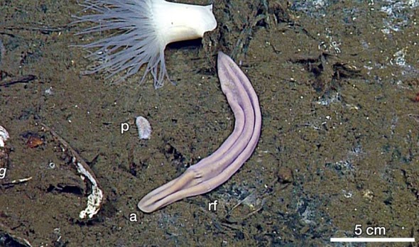
Figure 1: X. profunda in a clam field near a hydrothermal vent, Mexico. Abbreviations: a, anterior; rf, ring furrow; p, polynoid scaleworm. From Rouse et al. (2016), figure 1c. |
Going back to phylogenetics, the review by Nakano et al. (2015)[3] summarises the many complicated relationships Xenoturbella has entertained across the animal tree (figure 2). It was first described in 1949 as a flatworm in the phylum Platyhelminthes [A]. Morphological studies (admittedly without much to work with) then went on to identify it as a basal metazoan [B], basal bilaterian [C], deuterostome [D], bryozoan [E] and even a bivalve mollusc [F]. When molecular techniques arrived in 1997, a study comparing three genes surprisingly confirmed this bivalve affinity. But soon afterwards, another study using the very same genes found Xenoturbella to be a deuterostome again. It turned out that Xenoturbella could be identified as a bivalve if DNA was taken from the whole animal, but as a deuterostome if its gut was removed. Instead of finding its relatives, the 1997 study had inadvertently found its food source. Studies of other genes continued to find Xenoturbella in Deuterostomia, but in another jump across the animal tree, the first phylogenomic study (comparing essentially the entire genome) argued that it was actually a sister group to Acoelomorpha, another clade of simple wormy animals, so belonged right at the base of Bilateria.
First, they analysed the 13 mitochondrial proteins from all five species. The first phylogeny used the maximum likelihood method. This assumes that lineages evolve independently of each other, so are not closely related, so it should be well suited to deep phylogenetic scales such as this. It found Xenoturbella to be a sister group to the acoelomorphs, forming Xenacoelomorpha, a result which also appeared in all of their other analyses. So, Rouse et al. show very strong support for Xenacoelomorpha. The analysis then put Xenacoelomorpha with deuterostomes, as had been found in other mitochondrial studies[4], but with only weak support.
They then used the same mitochondrial data with another method, PhyloBayes. PhyloBayes distinguishes itself by accounting for site-specific amino acid or nucleotide preferences, so should be among the most accurate models of evolution[2]. It too returned Xenacoelomorpha within deuterostomes, but again with weak support. Rouse et al. suspect the low support in the two mitochondrial analyses might have been caused by their additional data from the new Xenoturbella species, or by using all 13 proteins instead of a selection. As noted above, there is reasonable evidence that higher taxon and character sampling usually reduce uncertainty in a phylogeny[5], so Xenoturbella being a deuterostome is looking unlikely.
Rouse et al. then moved to phylogenomics, selecting just X. profunda and X. bocki to represent Xenoturbella. Taking just two species for a genus seems efficient for phylogenies of this scale, but mitochondrial data for all Xenoturbella species was used in those analyses, and genome sequencing is now relatively cheap. It is interesting that Rouse et al. did not sequence the other species' genomes. Perhaps after submarine expeditions the budget was just a little too tight.
The first phylogenomic analysis performed was another maximum likelihood analysis, and it returned Xenacoelomorpha as the sister group to Nephrozoa, right at the base of Bilateria. This was the other dominant hypothesis, and it came with much better support. Several other maximum likelihood phylogenies testing for potential pitfalls, such as genes evolving at different speeds, were produced and all came to the same conclusion with similar support. The basal position looked strong.
Rouse et al. then used PhyloBayes again, this time with specific settings recommended by Philippe et al. (2011)[4] to avoid the dangers of long branch attraction (LBA). In their study, Philippe et al. had also performed a variety of phylogenetic methods and concluded that the position of Acoelomorpha, and Xenoturbella when they were found together, was biased by LBA. LBA is the tendency for distantly related taxa that branch near the base of a tree to appear closely related when they are not; they evolve similarities to each other by chance, and their lack of true close relatives means these similarities have no context[5]. Convergences can be wrongly interpreted as signs of relatedness. Philippe et al. argued that coincidental similarities between xenacoelomorphs and truly basal animals pulled them to the base of the tree. With LBA minimised, they recovered Xenacoelomorpha as deuterostomes. However, when Rouse et al. tried that method with their data Xenacoelomorpha came out in yet another new position, next to the deuterostomes' sister group Protostomia, with moderate support. They do not offer an explanation. However, published in the same issue as Rouse et al., Cannon et al. (2016)[1] also examined whether LBA affected Xenacoelomorpha, and found that it did not. So in hindsight, this PhyloBayes model may be an inappropriate correction after all, making Xenoturbella as a protostome unlikely.
Rouse et al. have succeeded in elevating our understanding of a mysterious genus. They discredit the placement of Xenoturbella in Deuterostomia, giving most (though not unanimous) support to placement at the base of Bilateria. While investigating the different methodologies that may have led to Xenoturbella's phylogenetic wandering, they found problems with phylogenies based on mitochondrial analyses, a common alternative to using whole genomes. This suggests that mitochondrial data might not be appropriate at deep phylogenetic scales. Here, however, it was very useful in confirming four new species to an enigmatic genus. Nakano et al.[3] were correct in suspecting that more Xenoturbella species were waiting to be found in the deep sea, and it is likely that more are waiting still.
References
[1] Johanna Taylor Cannon, Bruno Cossermelli Vellutini, Julian Smith, Fredrik Ronquist, Ulf Jondelius, and Andreas Hejnol. Xenacoelomorpha is the sister group to Nephrozoa. Nature, 530(7588):89?93, February 2016.
[2] Nicolas Lartillot, Thomas Lepage, and Samuel Blanquart. PhyloBayes 3: a Bayesian software package for phylogenetic reconstruction and molecular dating. Bioinformatics, 25(17):2286?2288, September 2009.
[3] Hiroaki Nakano. What is Xenoturbella? Zoological Letters, 1:22, 2015.
[4] Hervé Philippe, Henner Brinkmann, Richard R. Copley, Leonid L. Moroz, Hiroaki Nakano, Albert J. Poustka, Andreas Wallberg, Kevin J. Peterson, and Maximilian J. Telford. Acoelomorph flatworms are deuterostomes related to Xenoturbella. Nature, 470(7333):255?258, February 2011.
[5] Steven Poe. Evaluation of the Strategy of Long-Branch Subdivision to Improve the Accuracy of Phylogenetic Methods. Systematic Biology, 52(3):423?428, 2003.
[6] Greg W. Rouse, Nerida G. Wilson, Jose I. Carvajal, and Robert C. Vrijenhoek. New deep-sea species of Xenoturbella and the position of Xenacoelomorpha. Nature, 530(7588):94?97, February 2016.
Wednesday, 23 November 2016
Morphological phylogenetics
After weeks of travelling back and forth from another campus for computing and ecology, I'm now back at the NHM. An hour's commute each way never seemed so gentle.
This week is phylogenetics, starting in the same way the field did by completely ignoring DNA. We've been learning how to turn the morphological variation you see in organisms into hard facts that a computer can use to compare them, and through comparing many many features, suggest their evolutionary relationships. With molecular data you typically have a fixed number of characters: the base at this point in the sequence can only be A, C, T or G, for example. But there's a real art to choosing and defining morphological characters, as I found out yesterday.
In the practical session we were given a set of taxa to describe. My group came up with our set of characters and we ran the analysis and got a fairly nice cladogram, for something that took an hour. Most taxa had a clear set of relatives and you could trace the evolution of some of our characters back to common ancestors.
When I got back, I decided to ramp it up a notch, adding 16 taxa and changing some of the characters. I experimented with lots of different character arrangements, mostly unsuccessful. This not-brilliant cladogram was the result:
Did I mention the taxa were biscuits?
The methods used by morphological phylogenetics just assume there's an evolutionary relationship between the items you describe. One of my classmates apparently did one on screws.
So, to the science:
It seems most of the finger biscuits are closely related (yellow), and the two chocolate chip cookies have come out as sister taxa (green) as expected. Interestingly, the sandwich condition seems to have evolved multiple times, as it crops up all over the cladogram. The single-layer sandwich fingers (red) probably evolved the finger shape independently of the other fingers.
But, the tree has failed to recognise the close relationships between hobnobs and coated and uncoated digestives respectively; I feel these are unlikely to be convergences but represent true homology. It also failed to recover many deeper-level relationships, leaving a very large polytomy of most round biscuits. The single exception is the Viennese whirl as the basalmost biscuit (the jaffa cake is the outgroup). This is interesting, as the Viennese finger has come out as one of the most derived, so here the Viennese resemblance is likely to be convergent.
In conclusion, this analysis has revealed only a few useful characters with which biscuits can be classified, and these are still apparently subject to homoplasy. It is recommended that molecular methods be applied to offer a different perspective. Sadly, DNA extraction from biscuits is not an active field of research.
Sunday, 20 November 2016
Let the young make mistakes
This morning, a pair of woodpigeons dropped into the garden. It led me to much biological speculation.
I tried to take some photos:
Some readers may see a problem - these do not look like wood pigeons. There are a few differences probably beyond the scope of this questionable photo, like their big dark eyes and drab beaks, but it's clear that they lack the flashy collar. Wood pigeons have a bright white patch on the sides of their neck, with green iridescence around the back. This is because these birds are juveniles.
A typical reason for a juvenile to look different to an adult is sexual maturity: it makes no sense for a bird to have flashy colours to show off to potential mates if it's not interested in finding one, especially if the colours will interfere with its camouflage. This could definitely apply to these pigeons, even if pigeons are not known for camouflage (despite today's prevalence of grey concrete). But the adults aren't sexually dimorphic - males and females look the same - and flashy colours are almost always associated with just one sex. And watching them, it does seem to be females accepting or rejecting males rather than both having to look attractive. So what else could flashy colour be used for?
I've forgotten where, but I think I've read that another species of bird has a white patch near its head just like wood pigeons. It lives in groups, and the patch is a signal to its flockmates: when the bird suddenly takes off, the movement of the patch is very visible, so the rest of the flock can quickly follow. This makes sense if the first bird has spotted a predator, and it's in the interest of the flockmates to escape too. The white tails of deer and rabbits probably does the same job.
But importantly, the juveniles of this bird species also didn't have the patch. It only grows in with the adult feathers. So, a juvenile could take off and the other birds wouldn't necessarily feel the urge to follow. This might be selected for if juveniles are very prone to false positives: flying away from predators that don't actually exist. Ignoring the inexperienced younguns would save the adults (mostly family, if this speculation is going to make biological sense) lots of energy. Vervet monkeys, known for their different alarm calls that refer to different types of predator, will also usually ignore calls from juveniles. So, maybe these juvenile pigeons lacked the flashy collar not because they're pretty young to be searching for that kind of fun, but because it pays for their parents to ignore them. Anthropomorphise from that what you will.
Note: robins (photo two) also have very different colouration as juveniles. They're mottled brown, lacking the bright white and orange of adults. Adults of both sexes use their colours in competition over territories and can be very aggressive. Another unsourced factoid: 10% of robin deaths are caused by other robins. Lacking the colours might save the juveniles from being attacked by bigger birds before they need to find a territory. Maybe anthropomorphised robins would wait until voting age to reveal their political leanings.
I tried to take some photos:
The flash was on |
There's a robin |
The key picture |
Some readers may see a problem - these do not look like wood pigeons. There are a few differences probably beyond the scope of this questionable photo, like their big dark eyes and drab beaks, but it's clear that they lack the flashy collar. Wood pigeons have a bright white patch on the sides of their neck, with green iridescence around the back. This is because these birds are juveniles.
A typical reason for a juvenile to look different to an adult is sexual maturity: it makes no sense for a bird to have flashy colours to show off to potential mates if it's not interested in finding one, especially if the colours will interfere with its camouflage. This could definitely apply to these pigeons, even if pigeons are not known for camouflage (despite today's prevalence of grey concrete). But the adults aren't sexually dimorphic - males and females look the same - and flashy colours are almost always associated with just one sex. And watching them, it does seem to be females accepting or rejecting males rather than both having to look attractive. So what else could flashy colour be used for?
I've forgotten where, but I think I've read that another species of bird has a white patch near its head just like wood pigeons. It lives in groups, and the patch is a signal to its flockmates: when the bird suddenly takes off, the movement of the patch is very visible, so the rest of the flock can quickly follow. This makes sense if the first bird has spotted a predator, and it's in the interest of the flockmates to escape too. The white tails of deer and rabbits probably does the same job.
But importantly, the juveniles of this bird species also didn't have the patch. It only grows in with the adult feathers. So, a juvenile could take off and the other birds wouldn't necessarily feel the urge to follow. This might be selected for if juveniles are very prone to false positives: flying away from predators that don't actually exist. Ignoring the inexperienced younguns would save the adults (mostly family, if this speculation is going to make biological sense) lots of energy. Vervet monkeys, known for their different alarm calls that refer to different types of predator, will also usually ignore calls from juveniles. So, maybe these juvenile pigeons lacked the flashy collar not because they're pretty young to be searching for that kind of fun, but because it pays for their parents to ignore them. Anthropomorphise from that what you will.
Note: robins (photo two) also have very different colouration as juveniles. They're mottled brown, lacking the bright white and orange of adults. Adults of both sexes use their colours in competition over territories and can be very aggressive. Another unsourced factoid: 10% of robin deaths are caused by other robins. Lacking the colours might save the juveniles from being attacked by bigger birds before they need to find a territory. Maybe anthropomorphised robins would wait until voting age to reveal their political leanings.
Sunday, 6 November 2016
In the park II: what actually was in the park
Answers (highlight the text to see):
[A] Crow
[B] Nothing
[C] Nothing
[D] Crow
[E] Ring-neck parakeet
[F] Snake
Very surprising to see a reptile out on a cold autumn morning, but it wasn't moving much.
Yesterday I went to the other local park and the two highlights were a squirrel missing half its tail and a rat climbing out of a bin. But it was genuinely a very nice place.
![]() | |
Much more interesting than squirrels |
In the park I
In a rare burst of free time, I've been exploring the local green space. I went out early in the morning with my camera to see what non-squirrel creatures I could find.
Now, read carefully.
This is a game.
The combination of my photography and the sheer lack of wildlife makes this a great opportunity for spotting. What's the animal (if there is one)? Answers in the next post!
[A]
[B]
[C]
[D]
[E]
[F]
Now, read carefully.
This is a game.
The combination of my photography and the sheer lack of wildlife makes this a great opportunity for spotting. What's the animal (if there is one)? Answers in the next post!
[A]
[B]
[C]
[D]
[E]
[F]
Friday, 28 October 2016
Pretty patterns in R
The past two weeks has been all about programming in R, a language that's supposed to be the next great thing for statistics and computational biology. I'd already covered it in my last course and it was a source of great pain. This has been less pressured, but still challenging.
The highlight so far has been one section on building graphs. As far as I can tell, the beauty of R is that you can plot an awful lot of things in an awful lot of ways, but making even a simple line graph is waaaay more complicated than you'd imagine. This exercise was all about how to make a fairly complex graph from a matrix of data and then strip it all down to pretty quilt patterns. You should be able to paste this all into R (or RStudio) and run it, though I'd do it line-by-line to avoid formatting errors:
install.packages("ggplot2")
require(ggplot2)
require(reshape2) #reshape2 comes included with the basic R download
#We're going to plot a matrix of random values taken from a normal distribution #[0,1]. Because ggplot2 only accepts dataframes, we'll use reshape2 to 'melt' the matrix into a dataframe.
GenerateMatrix <- function(N){
M <- matrix(runif(N*N),N,N)
return(M)
}
GenerateMatrix(3) #The function fills a matrix with N*N random numbers from the uniform distribution. The matrix is N rows by N columns. So, the fill and size match.
M <- GenerateMatrix(10)
M #It's a 10 by 10 matrix
M[1:3,1:3] #Square brackets specify what you want to look at: this views just the first three rows and columns
Melt <- melt(M)
Melt[1:4,] #This object has many many rows (we see the first 4) and 3 columns (Var1, Var2 and value)
ggplot(Melt,aes(Var1,Var2,fill=value))+geom_tile() #The fill=value is what colours each tile according to its value. What a pretty diagram. But, we can make it prettier.
#Saving the plot as an object:
Plot <- ggplot(Melt,aes(Var1,Var2,fill=value))
Plot #It's just the blank plot; we're about to add another layer
Plot <- Plot+geom_tile()
Plot
#Removing the legend:
Plot2 <- Plot+theme(legend.position="none")
Plot2
#Removing all the rest:
Plot2 <- Plot+theme(legend.position="none",
panel.background=element_blank(),
axis.ticks=element_blank(),
panel.grid.major=element_blank(),
panel.grid.minor=element_blank(),
axis.text.x=element_blank(),
axis.title.x=element_blank(),
axis.text.y=element_blank(),
axis.title.y=element_blank())
Plot2 #It's now just a lovely blue grid
#Exploring the colours:
Plot3 <- Plot2
Plot3+scale_fill_gradient2()
Plot3+scale_fill_continuous(low="yellow",high="darkgreen")
Plot3+scale_fill_gradientn(colours=c("red","white","blue"))
Plot3+scale_fill_gradientn(colours=grey.colors(10)
Plot3+scale_fill_gradientn(colours=rainbow(10))
The highlight so far has been one section on building graphs. As far as I can tell, the beauty of R is that you can plot an awful lot of things in an awful lot of ways, but making even a simple line graph is waaaay more complicated than you'd imagine. This exercise was all about how to make a fairly complex graph from a matrix of data and then strip it all down to pretty quilt patterns. You should be able to paste this all into R (or RStudio) and run it, though I'd do it line-by-line to avoid formatting errors:
install.packages("ggplot2")
require(ggplot2)
require(reshape2) #reshape2 comes included with the basic R download
#We're going to plot a matrix of random values taken from a normal distribution #[0,1]. Because ggplot2 only accepts dataframes, we'll use reshape2 to 'melt' the matrix into a dataframe.
GenerateMatrix <- function(N){
M <- matrix(runif(N*N),N,N)
return(M)
}
GenerateMatrix(3) #The function fills a matrix with N*N random numbers from the uniform distribution. The matrix is N rows by N columns. So, the fill and size match.
M <- GenerateMatrix(10)
M #It's a 10 by 10 matrix
M[1:3,1:3] #Square brackets specify what you want to look at: this views just the first three rows and columns
Melt <- melt(M)
Melt[1:4,] #This object has many many rows (we see the first 4) and 3 columns (Var1, Var2 and value)
ggplot(Melt,aes(Var1,Var2,fill=value))+geom_tile() #The fill=value is what colours each tile according to its value. What a pretty diagram. But, we can make it prettier.
#Saving the plot as an object:
Plot <- ggplot(Melt,aes(Var1,Var2,fill=value))
Plot #It's just the blank plot; we're about to add another layer
Plot <- Plot+geom_tile()
Plot
#Removing the legend:
Plot2 <- Plot+theme(legend.position="none")
Plot2
#Removing all the rest:
Plot2 <- Plot+theme(legend.position="none",
panel.background=element_blank(),
axis.ticks=element_blank(),
panel.grid.major=element_blank(),
panel.grid.minor=element_blank(),
axis.text.x=element_blank(),
axis.title.x=element_blank(),
axis.text.y=element_blank(),
axis.title.y=element_blank())
Plot2 #It's now just a lovely blue grid
![]() | ||
Bathroom tiling |
#Exploring the colours:
Plot3 <- Plot2
Plot3+scale_fill_gradient2()
![]() |
Flowers for grandma |
Plot3+scale_fill_continuous(low="yellow",high="darkgreen")
![]() |
Aftermath of limeade explosion |
Plot3+scale_fill_gradientn(colours=c("red","white","blue"))
![]() |
Picnic blanket with a touch of chaos |
Plot3+scale_fill_gradientn(colours=grey.colors(10)
![]() |
Dodgy aerial connection (for anyone who remembers what an aerial is) |
Plot3+scale_fill_gradientn(colours=rainbow(10))
![]() |
My eyes are watering |
Saturday, 22 October 2016
Dinky interstitial phyla
The first week or so of this taxonomy course was a whizz through the tree of life, to get us all familiar with most types of organisms at the most basic level. One lecture managed to cover all fourteen phyla in the group of animals called Lophotrochozoa. For perspective, a phylum is a very deep level of organisation for animals that divides them by their basic body plan. Phyla vary hugely in how many species they contain, but we belong to the phylum Chordata, which contains every single vertebrate plus a few of our very close invertebrate relatives, like tunicates (sea squirts). The lecturer did a fine job of not running out of breath.
I'd met all of Lophotrochozoa briefly before, on my biology course, and two phyla stood out both times. I'd doodled them to try and remember what they looked like, and they are just adorable. Both contain exceedingly tiny animals that live between sand grains - that makes them 'interstitial', in the same way that animals that live on the sea floor are benthic. Meet Kinorhyncha and Gastrotricha respectively, in fabulous microscopy:
I know, they're like hairy sausage puppies with bowl-cuts and glitter, right? And in less fabulous Paint.NET (not to scale):
Or even in the Vesryn region:
References
Kinorhynch photo:
http://palaeos.com/metazoa/ecdysozoa/scalidophora/kinorhyncha.html
Gastrotrich photo:
https://tl.wikipedia.org/wiki/Talaksan:Gastrotrich.jpg
Screenshot adapted from Pokemon Zeta, a real gem of a game: https://www.reddit.com/r/pokemonzetaomicron/
I'd met all of Lophotrochozoa briefly before, on my biology course, and two phyla stood out both times. I'd doodled them to try and remember what they looked like, and they are just adorable. Both contain exceedingly tiny animals that live between sand grains - that makes them 'interstitial', in the same way that animals that live on the sea floor are benthic. Meet Kinorhyncha and Gastrotricha respectively, in fabulous microscopy:
I know, they're like hairy sausage puppies with bowl-cuts and glitter, right? And in less fabulous Paint.NET (not to scale):
![]() |
Or even in the Vesryn region:
References
Kinorhynch photo:
http://palaeos.com/metazoa/ecdysozoa/scalidophora/kinorhyncha.html
Gastrotrich photo:
https://tl.wikipedia.org/wiki/Talaksan:Gastrotrich.jpg
Screenshot adapted from Pokemon Zeta, a real gem of a game: https://www.reddit.com/r/pokemonzetaomicron/
Friday, 21 October 2016
Lattices and lettuces
I get hungry when working alone on the laptop and, if I'm going to get distracted by looking for something on the Internet, it will be food. I have a folder of recipes that's a direct result of this. It's a mixed blessing. Yesterday, I was working on a type of graph called lattice plots, and all I could think of were things like this1:
Not something that appeals to me nowadays (vegetarian), but a nice reminder of the times when somebody cooked for me. I've developed a morbid curiosity for highly processed, dirt cheap food, and was delighted to discover that someone has dedicated a blog to their exploration of it: www.barelyedible.co.uk It's a shame that most of the really impressive terrible food out there is meat-based as I'd love to join in.
In the meantime, I'll continue to look for yellow stickers in supermarkets on slightly less entertaining items. A really good recent find was this wonderful thing:
I'd eaten most of the big leaves a few days ago and the medium ones are quickly filling the gaps. There are too many plants in there for them to really grow healthily, but hopefully enough will hang on to feed one person a salad once in a while. I did keep another supermarket live pot on my desk for most of last year (Basil the basil, pronounced à la Sybil Fawlty) and three plants are just about still alive. Unlike most organisms, window plants like these succeed at being loveable even while you eat them.
References (as a good scientist I cite my sources)
1 http://www.mysupermarket.co.uk/tesco-price-comparison/Pies_And_Quiches/Tesco_Creamy_Chicken_Garlic_And_Mushroom_Lattice_Pie_550g.html
Monday, 17 October 2016
The French grass snake
My summer holiday this year involved a visit to the lovely French countryside. Best of all, a sun-baked barn in the place we were staying had become the final resting place of a very large grass snake (Natrix natrix), reduced to mostly skin and bone by the time we left. I toyed with the idea of taking the whole thing back but was wisely convinced to just snip the head off. I wrapped it in kitchen roll, taped it securely into part of an egg box and managed to get it home unscathed via three trains and a metro.
For the next few weeks I had the fun job of cleaning away the remaining mummified tissue and reassembling the skull and some cervical vertebrae. Some little palatal bones were damaged already, and tragically I snapped one with a vacuum cleaner and never found the end, but overall it's quite intact. I left the cervical ribs and some loose teeth out because they were SO fiddly, perhaps to try again one day. She* now sits proudly on my little nature shelf at home, in a perfectly-sized sputum collection vial never used for its intended purpose.
The holiday spot turned out to be a great place for snakes. I even managed to catch a little one by leaping at it Irwin-style, but immediately felt very sorry for it while it writhed around and ineffectually tried to bite me. It quickly went back into the wall. I hope the locals are kinder to their lovely reptiles.
*Wikipedia tells me that male grass snakes only get to about 50cm long, so at about a metre, mine was certainly a lady.
For the next few weeks I had the fun job of cleaning away the remaining mummified tissue and reassembling the skull and some cervical vertebrae. Some little palatal bones were damaged already, and tragically I snapped one with a vacuum cleaner and never found the end, but overall it's quite intact. I left the cervical ribs and some loose teeth out because they were SO fiddly, perhaps to try again one day. She* now sits proudly on my little nature shelf at home, in a perfectly-sized sputum collection vial never used for its intended purpose.
![]() |
(A, left lateral; B, dorsal; C, ventral; D, posterior; E, anterior views from a slightly shaky photographer) |
The holiday spot turned out to be a great place for snakes. I even managed to catch a little one by leaping at it Irwin-style, but immediately felt very sorry for it while it writhed around and ineffectually tried to bite me. It quickly went back into the wall. I hope the locals are kinder to their lovely reptiles.
*Wikipedia tells me that male grass snakes only get to about 50cm long, so at about a metre, mine was certainly a lady.
Tuesday, 11 October 2016
Why moths are attracted to lights
Yesterday was Plant Day, but first I thought I'd share something from last week's arthropod session. One of the entomologists revealed the answer to a something that has puzzled me for a long time: why moths are attracted to lights. It turns out to be a feature of moth navigation that works very well using natural light sources, but isn't well adapted dealing with human ones.
Younger moths navigate at night using light sources in the sky, like stars (older moths switch to using landmarks). Because the stars are so far away, the moth can fly in a straight line by maintaining a constant angle to where the star appears to be:
But artificial lights are significantly closer than stars, so when a moth flies at a constant angle to one it needs to keep compensating for how the light source 'moves', leading it into a spiral:
So, light pollution pulls moths away from their important nightly activities by making them fly in circles until the sun comes up. Do moths a favour by switching off unnecessary lights at night.
Saturday, 8 October 2016
Catching arthropods
So, lectures started on Thursday. Those and the ones next week will be an overview of the tree of life, getting us all up to speed on what things are before we start looking at more complicated things. There were some excellent speakers - you can really tell when a lecturer is enjoying themselves.
After whizzing through early vertebrates and fungi on Friday morning, we spent the afternoon learning how to catch terrestrial arthropods. This involved swinging nets and beating trees and digging holes around the NHM wildlife garden, to the amusement of passing visitors. The soil samples were taken back by one of the entomologists to be processed over the weekend: you can separate critters from crud by pouring the samples into a funnel and shining a lamp over the top. The animals normally move away from light (they show negative phototaxis), so they gradually crawl downwards and fall into a collecting pot.
We could see the things beaten out of trees straight away - indeed, a few flew off as soon as they hit the collecting sheet. It was great to have experts on hand to identify them, though by the end of the session one was suffering after pootering up lots of hemipterans (true bugs) that released unpleasant fumes. One of the most interesting creatures was another hemipteran, probably a thread-legged bug (Emesinae) according to a quick Internet search. It looked like a tiny tiny stick insect, about 1.5 centimetres long, but had front legs just like a mantis.
However, the highlight of the afternoon was near the beginning, when we were standing in the meadow for a long while watching a trap being set up. All of a sudden, someone noticed an inch-long common frog on their shoe. Soon we'd seen half a dozen of them hopping around in the grass, and everyone became very conscious of where they were putting their feet. The shoe frog stayed safe on high ground until we'd gone.
Thursday, 6 October 2016
Beating biology
Today I was waiting at a crossing with a four-year old girl and her carer.
Girl: I don't want to marry a man.
Lady: Why's that?
Girl: I never want to have babies. That's why I want to marry a lady.
Lady: You know, if you marry a lady you can still adopt babies.
*Girl's face falls*
Girl: I don't want to marry a man.
Lady: Why's that?
Girl: I never want to have babies. That's why I want to marry a lady.
Lady: You know, if you marry a lady you can still adopt babies.
*Girl's face falls*
Wednesday, 5 October 2016
Dinosaurs at the NHM
Not to brag, but my course is mostly being taught in London's Natural History Museum. In between induction things this week, I've been taking a quick look around the galleries, partly to try and learn my way around but mostly because museums are fantastic and the NHM has a stellar reputation.
Most things were great, I'd like to make that clear. But the dinosaur gallery, the most anticipated part for me, was a bit disappointing. On the whole it seemed very well designed, and there were some fabulous mounts and no glaring errors in the text. There were lots of touchables and the school party in with me was having a great time. Unfortunately, every few minutes I'd notice something out of date, and with the exception of a note about feathered dromaeosaurs I didn't see any corrections.
The theropods were most obvious: there were several pronated hands*, though not universally, and many an upright body in the images. I quite enjoy retro palaeoart (I'm pretty sure a print of this was in there, carrying on the timeless trope of Deinonychus vs. Tenontosaurus: http://www.johnsibbick.com/library/display.asp?page=15&product=D15), but not when it's presented alongside up-to-date information with no caveat. There was also a case of nice shoebox-sized dinosaur sculptures, but again, most were totally vintage. There was even a humpy Stegosaurus that could have passed for a 1975 Invicta**.
In a shot at redemption that somewhat missed the mark, there were two animatronic Deinonychus near the end, with the note about feathers, that were indeed feathered. Unfortunately, I don't think they looked great. Most of the body had a short, shaggy coat that looked more like fur, with a token row of vaned feathers along each forearm and bare hands. I'm not basing this on primary sources, but I was under the impression that feathers in dromaeosaurs likely covered the hands to form a pretty clear wing, even if they didn't grow from the fingers. And, sadly, the hands were pronated.
Unfortunately, as I've grown more knowledgeable about dinosaurs I've just gotten more thorough in nit-picking. It was a fun exhibition with some solid science and great displays, just lacking in corrective signage. Hopefully it will get an update eventually, and the museum can help catch the public up with scientists.
I'd like to note that (at least on a quick view) I have nothing bad to say about the other galleries, and palaeontology is a particularly fast-changing thing. I have yet to see a dinosaur exhibition I can't pick faults with. Guess I'd better make one one day!
*The eminently readable Dave Hone explains the pronation problem: https://archosaurmusings.wordpress.com/2009/05/27/theropods-are-clappers-not-slappers/
**My summer guilty-pleasure reading: http://dinotoyblog.com/2010/07/22/stegosaurus-invicta/
Another footnote: I have the inevitable start-of-term cold today so might have been feeling less charitable than usual.
Early observations
So far this week, I have...
- Seen my first ring-necked parakeets, flying overhead. They sounded like a cross between a squeaky toy and a particularly irritable sparrow.
- Encountered my first manspreader on the Tube. I was bunched up against his right thigh. Thanks.
- Sat in class doing nothing for half an hour when we were supposed to have an IT induction, because IT didn't know we were supposed to have an IT induction.
- Sat in a lecture theatre watching colleagues play charades for half an hour when we were supposed to have a safety induction, because we'd all been given the wrong room for the safety induction.
- Got lots of exercise in a futile search for said safety induction.
- Lost faith in the organisational ability of the university.
Grey snot
Hello world,
I've just moved to London to start a course on taxonomy, the area of biology that deals with naming and classifying things. I'm hoping it will suit my joint interests of organising things and dinosaurs.
It's been a long couple of days; I moved into my new rented room on Sunday afternoon, then Monday was a 13-hour day of going through induction and shopping for things I'd forgotten. But I've been impressed with the efficiency of the Tube so far, and walking through central London is entertaining, if a bit surreal.
I'm going to try and use this blog to keep a record of what's sure to be a strange twelve months. It's my first permanent move away from home (the holidays during my undergraduate degree made me feel like a part-timer) and hopefully the first major step towards Dream Job. I'd love to be a natural history curator. I just hope museums will still have paid staff by the time I qualify!
Final thought: since moving down I've produced grey snot. There's no air pollution like London air pollution.
Subscribe to:
Posts (Atom)