Tuesday, 6 December 2016

Into the laboratory II

Day two of three was split in half.
1. Another method of DNA extraction, using the two cricket legs from Friday
2. PCR (https://www.youtube.com/watch?v=mvvP90Cpdfc)

So, last time we extracted DNA we ground up samples and soaked them in various chemicals, then used centrifuging to separate the liquid containing the DNA from the rest of the mixture. This time, we used a method which keeps the sample more or less intact. The museum does this to analyse its collection specimens, for which grinding up is discouraged. The cricket legs had been stored in a solution containing enzymes over the weekend. These released small amounts of DNA into the solution without changing the morphology of the leg, so if we really wanted to, we could put the legs out on display and nobody would know what we'd done.

The downside of this method is that it can be difficult to get enough DNA out to work with. Fortunately, my two leg samples turned out to be fine. After washing away lots of the protein and other stuff, I ran them through a NanoDrop again which showed that DNA was present and fairly uncontaminated, and then through another instrument called a Qubit which quantified the amount more accurately. I do not remember the number but I'm sure the average reader won't mind.

So, with DNA from both extraction attempts, it was time for PCR. The ultimate goal of most DNA extraction (though not ours because we only had three days) is to sequence it, and sequencing needs relatively large amounts of very pure DNA, of just the parts you want to sequence. For example, the cricket leg was probably covered in bacteria, and bacterial DNA probably got into the tube, but I wouldn't be interested in sequencing the bacterias' DNA. The solution, to purifying and increasing quantity, is the polymerase chain reaction: PCR.

Without going into terrifying bichemistry, PCR takes advantage of how DNA naturally replicates. Recall that DNA is ladder-shaped, with two long backbones connected by many rungs. Each 'rung' is actually two bases, the parts that code the information. The sequence of bases is equivalent to the strings of 0s and 1s that define a computer program. There are four types of base in DNA: A, T, C and G.
 
To copy itself, DNA splits lengthways, separating the backbones, each of which takes the bases it's attached to. Each side can then collect new bases and build a new second backbone. The key is that each base can only join to one other base: base A joins with T, and C joins with G. So, when the missing half of the molecule is reconstructed it forms a perfect (in theory - mistakes do happen and we call them mutations) copy of the one that was lost.

(See that the incoming bases bring a bit of backbone with them)


PCR is essentially this process in a test tube, with one important addition: rather than allowing the entire molecule to replicate, it starts and stops replication in specific places. So, you can have a whole genome in the tube but only replicate the gene you're interested in. There are so many copies of that gene that by the end, the concentration of the original DNA molecules is nearly zero, and the sample of that gene is extremely pure.

I spent most of the afternoon preparing the PCR tubes. Every one (of twenty) needed the right amount of PCR 'mastermix', which provides the ingredients for DNA replication. The quantities are so very tiny, even an extra half a microlitre could stop the reaction from happening at all. The pipettes are very clever but you still need to be very organised. You can't just look at the amount in the tube to see whether you added the last ingredient! Finally, I added my samples of DNA to their own individual tubes and loaded them into the thermocycler. This machine would carefully control the temperature, stopping and starting the PCR reaction while I left to catch my train. The results would have to wait until tomorrow...

Monday, 5 December 2016

Into the laboratory I

After a lot more theory and computing, we've started what's known in the business as wet lab work: chemicals and pipetting and Eppendorf tubes. We're hoping to put our new-found knowledge about turning DNA sequence data into phylogenetic trees to use. Day one of three was about extracting DNA from organisms.

First, I took the tiniest little palm-sized mortar and pestle and ground up the samples. These were two bits of mystery leaf (probably spinach and coriander) and two crickets. The leaves were very simple, but the crickets had to be disassembled.

Apologies to entomologists who enjoy anatomical correctness
A) One back leg set aside for another analysis next week
B) All other legs removed
C) Head removed (there's apparently something in the head which interferes with the chemistry of the DNA-replicating reaction)
D) Guts squeezed out and the outside of the abdomen discarded

It was a mercy that the crickets didn't smell too strong. But squeezing out the innards of the big one was utterly repulsive.

With each ground-up sample in its own little tube, I then let them soak in various chemicals to separate the DNA from the rest of the stuff. To then physically separate the different materials, the tubes were spun in a centrifuge. This little machine (about half the size of a microwave) contains something like a tiny roulette wheel, with slots for tubes around the outside. Their bottoms face outwards. As it spins (super fast; making use of centrifugal force), the heaviest material in the tubes is forced to the very bottom. The next-lightest material forms a distinct layer on top of that, then the next-lightest material, and so on. Here, the DNA was lightest, so I had a nice big layer of the liquid holding the DNA at the top.


This liquid was removed to its own new tube, and the rest discarded. There's a real skill to using the pipettes. They're very well crafted for moving tiny amounts of liquid around, but the liquid sure can move fast. Despite having some experience with them I did manage to spray myself with chloroform when my pipette unexpectedly squirted.

So, with a reasonably purified DNA suspension, the next step was to get rid of the suspension and replace it with another liquid. I honestly cannot remember why. But, the first one was a carcinogen and I've already proven liable to spray it over me.

I first added something which made the DNA gather together into a visible cloudy mass. Another centrifuge packed this mass down into what's referred to as a pellet, though in practice it's more of a tiny whiteish smear.


I sucked out all of the liquid I could and then rinsed my tiny whiteish smears in ethanol to dissolve anything else away. I did suck out most of the ethanol too, but because it evaporates so easily the final drying out was done by a much easier hot surface. Finally, I added the presumably non-carcinogenic solvent and watched my tiny whiteish smears dissolve away again.

The last task of the day was to take out a tiny tiny amount from each of my tiny tubes of tiny sample and put them through a machine called a NanoDrop. This uses lasers to quite literally see how much DNA there is and how much contaminant is left. It's not the most accurate, but gave all of my samples a tick of approval, which was a pleasant surprise. I've done this sort of procedure once before, back in the depths of undergrad, and few of us managed to get it to work at all. So, day one for me was a success.

Wet lab is turning out to be less frightening than I thought.


An article on surprisingly troublesome wormy things

There have been two key differences so far between this course and my undergraduate. Compared to a typical autumn term back then, this has had:
1. Two vs. zero group presentations, which were painful but not quite as horrific as feared.
2. One vs. eight essays to write. But I'm probably spending the equivalent time on extra travel!

Anyhow, I present this term's finished essay. It's designed to be like Nature's News & Views articles, which talk about interesting new papers in a relatively friendly way, accessible to scientists from other disciplines and the interested amateur.

______________________________________________________________________________________

Xenoturbella and phylogenetic wanderlust

Phylogenetics, the evolutionary relationships of organisms, is crucial to understanding them fully. How else could we appreciate how, for example, vertebrate fins turned into limbs and back again? Finding the close relatives of an organism can also help us reconstruct its ancestors. For many areas in the animal tree of life, molecular analysis has complemented and clarified phylogenetic trees built on morphology alone, and we can be reasonably confident that the relationships we infer are real. Unfortunately, there are some problematic taxa whose placement, despite molecular analysis, is still a mystery. Xenoturbella, a genus of small benthic worms, is one. The debate over what type of animal Xenoturbella is has recently condensed to two hypotheses: does it belong at the base of Deuterostomia, potentially informing on the ancestor of vertebrates and our close relatives, or at the base of Bilateria, informing on the ancestor of Deuterostomes and a whole lot more? In their 2016 paper, Rouse et al.[6] raise the known number of Xenoturbella species to five, assess phylogenetic methods suggested for such deep timescales and present results that strongly suggest Xenoturbella belongs at the base of Bilateria... or maybe not. The puzzle is not quite solved yet.

Prior to their paper, Xenoturbella was represented by just two species, X. bocki and X. westbladi, both found only off the west coast of Sweden. As predicted by Nakano et al.[3], more Xenoturbella were waiting to be found in the deep sea, and Rouse et al. collected multiple specimens from three sites in the east Pacific. According to their mitochondrial genes, they represented four new species: X. monstrosa, X. profunda (figure 1), X. hollandorum and the delightfully named X. churro. The attentive reader may have noticed a mismatch in numbers; the mitochondrial genes of the original Xenoturbella species were similar enough that Rouse et al. treat them as synonyms, so X. westbladi has been cast out to taxonomic purgatory. Nevertheless, the diversity of the genus has been significantly increased. Having more species allows greater taxon sampling in phylogenetic analyses, which may improve results by reducing the impact of errors like long branch attraction (see below), though it has the potential to cause other problems[5]. However, more Xenoturbella species is probably not relevant to its position within the entire animal phylogeny; at such a deep scale the differences between species in the same genus are essentially zero.
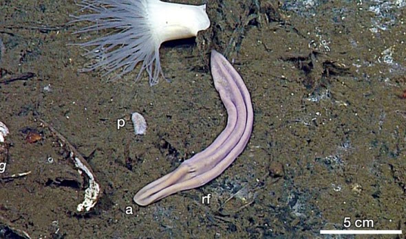
Figure 1: X. profunda in a clam field near a hydrothermal vent, Mexico. Abbreviations: a, anterior; rf, ring furrow; p, polynoid scaleworm. From Rouse et al. (2016), figure 1c.
 Placing Xenoturbella using morphology has been dificult because they are very simple animals, lacking a centralised nervous system, coelom, excretory or reproductive organs. They glide across surfaces using ventral cilia. Quite how they find their way is unknown: an organ near the head-end may be a balance-sensing statocyst, and a sensory function has also been suggested for the furrows that encircle the body and extend down the sides, but neither are confirmed[3]. Rouse et al. pickled their new specimens shortly after collection; it would be interesting if return expeditions could perform behavioural studies on the new species.

Going back to phylogenetics, the review by Nakano et al. (2015)[3] summarises the many complicated relationships Xenoturbella has entertained across the animal tree (figure 2). It was first described in 1949 as a flatworm in the phylum Platyhelminthes [A]. Morphological studies (admittedly without much to work with) then went on to identify it as a basal metazoan [B], basal bilaterian [C], deuterostome [D], bryozoan [E] and even a bivalve mollusc [F]. When molecular techniques arrived in 1997, a study comparing three genes surprisingly confirmed this bivalve affinity. But soon afterwards, another study using the very same genes found Xenoturbella to be a deuterostome again. It turned out that Xenoturbella could be identified as a bivalve if DNA was taken from the whole animal, but as a deuterostome if its gut was removed. Instead of finding its relatives, the 1997 study had inadvertently found its food source. Studies of other genes continued to find Xenoturbella in Deuterostomia, but in another jump across the animal tree, the first phylogenomic study (comparing essentially the entire genome) argued that it was actually a sister group to Acoelomorpha, another clade of simple wormy animals, so belonged right at the base of Bilateria.

Figure 2: A simplified metazoan phylogeny showing the various placements of Xenoturbella. [A], in Platyhelminthes (Protostomia); [B], at the base of Metazoa; [C], at the base of Bilateria; [D], in Deuterostomia; [E], in Bryozoa and [F] in Mollusca (both Protostomia).
Despite subsequent work, basal-Deuterostomia and basal-Bilateria are still competing hypotheses. It has been suggested that the phylogenetic methods used are a source of conflict between them, which is quite reasonable. Because we cannot see back in time, it is usually impossible to know whether reconstructed phylogenetic relationships are true, making phylogenetics a field of better or worse hypotheses rather than facts. To address some potential methodological issues, Rouse et al. used several different methods to fit Xenoturbella into the animal tree and compared the results.

First, they analysed the 13 mitochondrial proteins from all five species. The first phylogeny used the maximum likelihood method. This assumes that lineages evolve independently of each other, so are not closely related, so it should be well suited to deep phylogenetic scales such as this. It found Xenoturbella to be a sister group to the acoelomorphs, forming Xenacoelomorpha, a result which also appeared in all of their other analyses. So, Rouse et al. show very strong support for Xenacoelomorpha. The analysis then put Xenacoelomorpha with deuterostomes, as had been found in other mitochondrial studies[4], but with only weak support.

They then used the same mitochondrial data with another method, PhyloBayes. PhyloBayes distinguishes itself by accounting for site-specific amino acid or nucleotide preferences, so should be among the most accurate models of evolution[2]. It too returned Xenacoelomorpha within deuterostomes, but again with weak support. Rouse et al. suspect the low support in the two mitochondrial analyses might have been caused by their additional data from the new Xenoturbella species, or by using all 13 proteins instead of a selection. As noted above, there is reasonable evidence that higher taxon and character sampling usually reduce uncertainty in a phylogeny[5], so Xenoturbella being a deuterostome is looking unlikely.

Rouse et al. then moved to phylogenomics, selecting just X. profunda and X. bocki to represent Xenoturbella. Taking just two species for a genus seems efficient for phylogenies of this scale, but mitochondrial data for all Xenoturbella species was used in those analyses, and genome sequencing is now relatively cheap. It is interesting that Rouse et al. did not sequence the other species' genomes. Perhaps after submarine expeditions the budget was just a little too tight.

The first phylogenomic analysis performed was another maximum likelihood analysis, and it returned Xenacoelomorpha as the sister group to Nephrozoa, right at the base of Bilateria. This was the other dominant hypothesis, and it came with much better support. Several other maximum likelihood phylogenies testing for potential pitfalls, such as genes evolving at different speeds, were produced and all came to the same conclusion with similar support. The basal position looked strong.

Rouse et al. then used PhyloBayes again, this time with specific settings recommended by Philippe et al. (2011)[4] to avoid the dangers of long branch attraction (LBA). In their study, Philippe et al. had also performed a variety of phylogenetic methods and concluded that the position of Acoelomorpha, and Xenoturbella when they were found together, was biased by LBA. LBA is the tendency for distantly related taxa that branch near the base of a tree to appear closely related when they are not; they evolve similarities to each other by chance, and their lack of true close relatives means these similarities have no context[5]. Convergences can be wrongly interpreted as signs of relatedness. Philippe et al. argued that coincidental similarities between xenacoelomorphs and truly basal animals pulled them to the base of the tree. With LBA minimised, they recovered Xenacoelomorpha as deuterostomes. However, when Rouse et al. tried that method with their data Xenacoelomorpha came out in yet another new position, next to the deuterostomes' sister group Protostomia, with moderate support. They do not offer an explanation. However, published in the same issue as Rouse et al., Cannon et al. (2016)[1] also examined whether LBA affected Xenacoelomorpha, and found that it did not. So in hindsight, this PhyloBayes model may be an inappropriate correction after all, making Xenoturbella as a protostome unlikely.

Rouse et al. have succeeded in elevating our understanding of a mysterious genus. They discredit the placement of Xenoturbella in Deuterostomia, giving most (though not unanimous) support to placement at the base of Bilateria. While investigating the different methodologies that may have led to Xenoturbella's phylogenetic wandering, they found problems with phylogenies based on mitochondrial analyses, a common alternative to using whole genomes. This suggests that mitochondrial data might not be appropriate at deep phylogenetic scales. Here, however, it was very useful in confirming four new species to an enigmatic genus. Nakano et al.[3] were correct in suspecting that more Xenoturbella species were waiting to be found in the deep sea, and it is likely that more are waiting still.


References
[1] Johanna Taylor Cannon, Bruno Cossermelli Vellutini, Julian Smith, Fredrik Ronquist, Ulf Jondelius, and Andreas Hejnol. Xenacoelomorpha is the sister group to Nephrozoa. Nature, 530(7588):89?93, February 2016.
[2] Nicolas Lartillot, Thomas Lepage, and Samuel Blanquart. PhyloBayes 3: a Bayesian software package for phylogenetic reconstruction and molecular dating. Bioinformatics, 25(17):2286?2288, September 2009.
[3] Hiroaki Nakano. What is Xenoturbella? Zoological Letters, 1:22, 2015.
[4] Hervé Philippe, Henner Brinkmann, Richard R. Copley, Leonid L. Moroz, Hiroaki Nakano, Albert J. Poustka, Andreas Wallberg, Kevin J. Peterson, and Maximilian J. Telford. Acoelomorph flatworms are deuterostomes related to Xenoturbella. Nature, 470(7333):255?258, February 2011.
[5] Steven Poe. Evaluation of the Strategy of Long-Branch Subdivision to Improve the Accuracy of Phylogenetic Methods. Systematic Biology, 52(3):423?428, 2003.
[6] Greg W. Rouse, Nerida G. Wilson, Jose I. Carvajal, and Robert C. Vrijenhoek. New deep-sea species of Xenoturbella and the position of Xenacoelomorpha. Nature, 530(7588):94?97, February 2016.

Wednesday, 23 November 2016

Morphological phylogenetics

After weeks of travelling back and forth from another campus for computing and ecology, I'm now back at the NHM. An hour's commute each way never seemed so gentle.

This week is phylogenetics, starting in the same way the field did by completely ignoring DNA. We've been learning how to turn the morphological variation you see in organisms into hard facts that a computer can use to compare them, and through comparing many many features, suggest their evolutionary relationships. With molecular data you typically have a fixed number of characters: the base at this point in the sequence can only be A, C, T or G, for example. But there's a real art to choosing and defining morphological characters, as I found out yesterday.

In the practical session we were given a set of taxa to describe. My group came up with our set of characters and we ran the analysis and got a fairly nice cladogram, for something that took an hour. Most taxa had a clear set of relatives and you could trace the evolution of some of our characters back to common ancestors.

When I got back, I decided to ramp it up a notch, adding 16 taxa and changing some of the characters. I experimented with lots of different character arrangements, mostly unsuccessful. This not-brilliant cladogram was the result:


Did I mention the taxa were biscuits?

The methods used by morphological phylogenetics just assume there's an evolutionary relationship between the items you describe. One of my classmates apparently did one on screws.

So, to the science:

It seems most of the finger biscuits are closely related (yellow), and the two chocolate chip cookies have come out as sister taxa (green) as expected. Interestingly, the sandwich condition seems to have evolved multiple times, as it crops up all over the cladogram. The single-layer sandwich fingers (red) probably evolved the finger shape independently of the other fingers.

But, the tree has failed to recognise the close relationships between hobnobs and coated and uncoated digestives respectively; I feel these are unlikely to be convergences but represent true homology. It also failed to recover many deeper-level relationships, leaving a very large polytomy of most round biscuits. The single exception is the Viennese whirl as the basalmost biscuit (the jaffa cake is the outgroup). This is interesting, as the Viennese finger has come out as one of the most derived, so here the Viennese resemblance is likely to be convergent.

In conclusion, this analysis has revealed only a few useful characters with which biscuits can be classified, and these are still apparently subject to homoplasy. It is recommended that molecular methods be applied to offer a different perspective. Sadly, DNA extraction from biscuits is not an active field of research.

Sunday, 20 November 2016

Let the young make mistakes

This morning, a pair of woodpigeons dropped into the garden. It led me to much biological speculation.


I tried to take some photos:

The flash was on

There's a robin

The key picture

Some readers may see a problem - these do not look like wood pigeons. There are a few differences probably beyond the scope of this questionable photo, like their big dark eyes and drab beaks, but it's clear that they lack the flashy collar. Wood pigeons have a bright white patch on the sides of their neck, with green iridescence around the back. This is because these birds are juveniles.

A typical reason for a juvenile to look different to an adult is sexual maturity: it makes no sense for a bird to have flashy colours to show off to potential mates if it's not interested in finding one, especially if the colours will interfere with its camouflage. This could definitely apply to these pigeons, even if pigeons are not known for camouflage (despite today's prevalence of grey concrete). But the adults aren't sexually dimorphic - males and females look the same - and flashy colours are almost always associated with just one sex. And watching them, it does seem to be females accepting or rejecting males rather than both having to look attractive. So what else could flashy colour be used for?

I've forgotten where, but I think I've read that another species of bird has a white patch near its head just like wood pigeons. It lives in groups, and the patch is a signal to its flockmates: when the bird suddenly takes off, the movement of the patch is very visible, so the rest of the flock can quickly follow. This makes sense if the first bird has spotted a predator, and it's in the interest of the flockmates to escape too. The white tails of deer and rabbits probably does the same job.

But importantly, the juveniles of this bird species also didn't have the patch. It only grows in with the adult feathers. So, a juvenile could take off and the other birds wouldn't necessarily feel the urge to follow. This might be selected for if juveniles are very prone to false positives: flying away from predators that don't actually exist. Ignoring the inexperienced younguns would save the adults (mostly family, if this speculation is going to make biological sense) lots of energy. Vervet monkeys, known for their different alarm calls that refer to different types of predator, will also usually ignore calls from juveniles. So, maybe these juvenile pigeons lacked the flashy collar not because they're pretty young to be searching for that kind of fun, but because it pays for their parents to ignore them. Anthropomorphise from that what you will.


Note: robins (photo two) also have very different colouration as juveniles. They're mottled brown, lacking the bright white and orange of adults. Adults of both sexes use their colours in competition over territories and can be very aggressive. Another unsourced factoid: 10% of robin deaths are caused by other robins. Lacking the colours might save the juveniles from being attacked by bigger birds before they need to find a territory. Maybe anthropomorphised robins would wait until voting age to reveal their political leanings.

Sunday, 6 November 2016

In the park II: what actually was in the park

Answers (highlight the text to see):
[A] Crow
[B] Nothing
[C] Nothing
[D] Crow
[E] Ring-neck parakeet
[F] Snake

Very surprising to see a reptile out on a cold autumn morning, but it wasn't moving much.

Yesterday I went to the other local park and the two highlights were a squirrel missing half its tail and a rat climbing out of a bin. But it was genuinely a very nice place.


Much more interesting than squirrels

In the park I

In a rare burst of free time, I've been exploring the local green space. I went out early in the morning with my camera to see what non-squirrel creatures I could find.

Now, read carefully.

This is a game.

The combination of my photography and the sheer lack of wildlife makes this a great opportunity for spotting. What's the animal (if there is one)? Answers in the next post!

[A]


[B]

[C]

[D]
[E]

[F]