Note: I've pasted in text, which has resulted in some amusing font issues. I haven't worked out how to fix them. Such is life.


The spotted green
pigeon, Caloenas maculata, is
extinct. It was described in 1783 based on two specimens.
One was collected by
Sir Joseph Banks, one by General Thomas Davies. But then the Banks specimen was lost...

... Leaving us with just one. And it lives in the World Museum...

... in Liverpool. In fact, its other common name is the Liverpool pigeon.
Unfortunately, nobody knows where either specimen originally came from.

But
both Banks and Davies did most of their collecting in Oceania, so probably from
there.

Here's a
reconstruction. It's a fairly standard tropical pigeon. It could definitely fly.
Apparently its short, rounded wings suggest that it lived on islands. But to
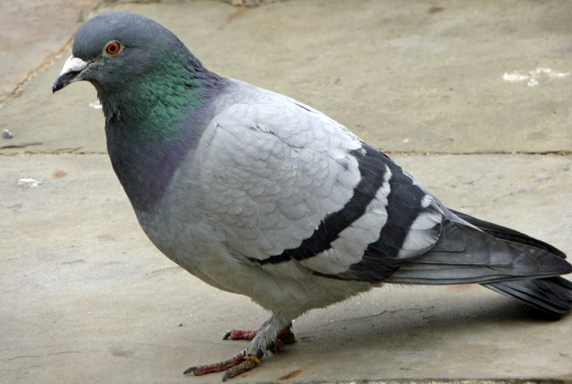
infer more about it, it would be helpful to know what its closest relatives are.

Unfortunately,
the spotted green has rather jumped around the pigeon tree. The big group
containing European pigeons is Columbinae at the top. Raphinae has the huge
diversity of tropical pigeons.
Based
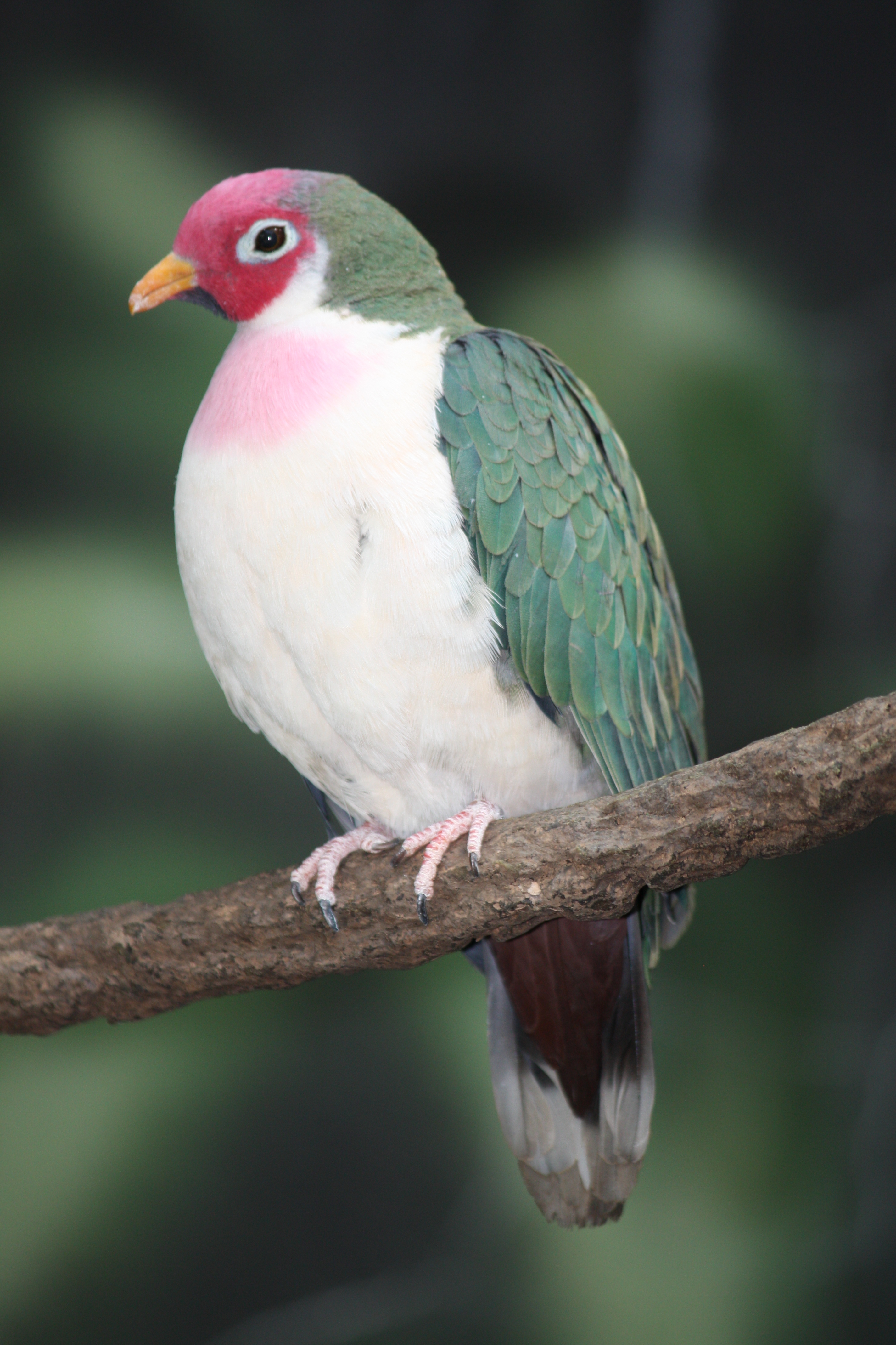
on morphology, the spotted green has been placed somewhere in Ptilinopini,
sister to the imperial, mountain or fruit pigeons.
What
technique can help resolve taxonomic questions based only on morphology? DNA
analysis! Which is where Heupink and colleagues come in.

The
team managed to extract DNA from two feathers. Because that one remaining
specimen is a couple of hundred years old, they used ancient DNA techniques,
which included lots of contamination precautions and lots of controls.


There
were two key improvements, both on the final step of DNA extraction, when you
bind the DNA to a membrane and wash away impurities.

First,
they used a binding buffer with more isopropanol. This encouraged the shorter
DNA molecules to actually bind. Another group had already tried this and it
worked for them. The
second thing was to wash with the buffer in the kit multiple times, and also
with phenol. This removed more PCR inhibitors, quantitatively and
qualitatively.
So
finally...

They
succeeded in getting detectable, amplifiable DNA! Unfortunately...

The
fragments were super short. Like less than 30 base pairs long.

The
quantity was really low.

And
for one of the two feathers, the DNA was super fragmented too.

But
at least they had DNA. The
idea had been to analyse a barcode from the mitochondrial 12S gene. Their
reference dataset was over a hundred 12S sequences from pigeons, downloaded from GenBank.
But the spotted green fragments were too short for a typical barcode...
But the spotted green fragments were too short for a typical barcode...

... so the team
designed three
mini-barcodes instead. These
turned out to work really well. They sequenced successfully and consistently,
and...

... a
BLAST search said
that the sequences came from an unknown pigeon, which was exactly what they
wanted.

Using
the mini-barcodes, and all of the reference sequences, they built a maximum
likelihood tree for each feather, and they were pretty much identical, which
was a relief. All
but one of the other taxa ended up where they were expected to - the pigeon
tree isn’t completely stable yet, but this was consistent with previous
findings. But the only support metric they’ve used there is bootstrap, which
sticks in my mind from the cladistics course as something you shouldn’t rely on
too hard. It would have been nice to see another measure there too.

But
anyway, This is a zoom in on one of them. the spotted green pigeon comes out
closest to the Nicobar with high bootstrap support (it's out of 100), for what it’s worth. They
are very similar after all.


There
was an average of 90.5% pairwise identity between the spotted green and the
Nicobar. This translated to about 0.11 substitutions per site. The average
difference within species was 0.01 substitutions per site, and the average
difference between species of the same genus was 0.09.

T-tests
showed that the spotted-Nicobar average was significantly different from
within-species, but not from within-genus.

So, the spotted green
shares a genus with the Nicobar pigeon but is different enough to be its own
species.


The Kanaka pigeon is
extinct, and is only known from a handful of subfossils found on New Caledonia
and Tonga: hot, humid, beautiful places that turn DNA into soup. The boundaries of ancient
DNA keep being pushed, but it’s not there yet.

Also, the Nicobar is
bigger than the spotted green, and the Kanaka is bigger than the Nicobar, so we
can be pretty sure it’s not the same species as the spotted green. Leaving it
out doesn’t increase confusion over what the spotted green is.

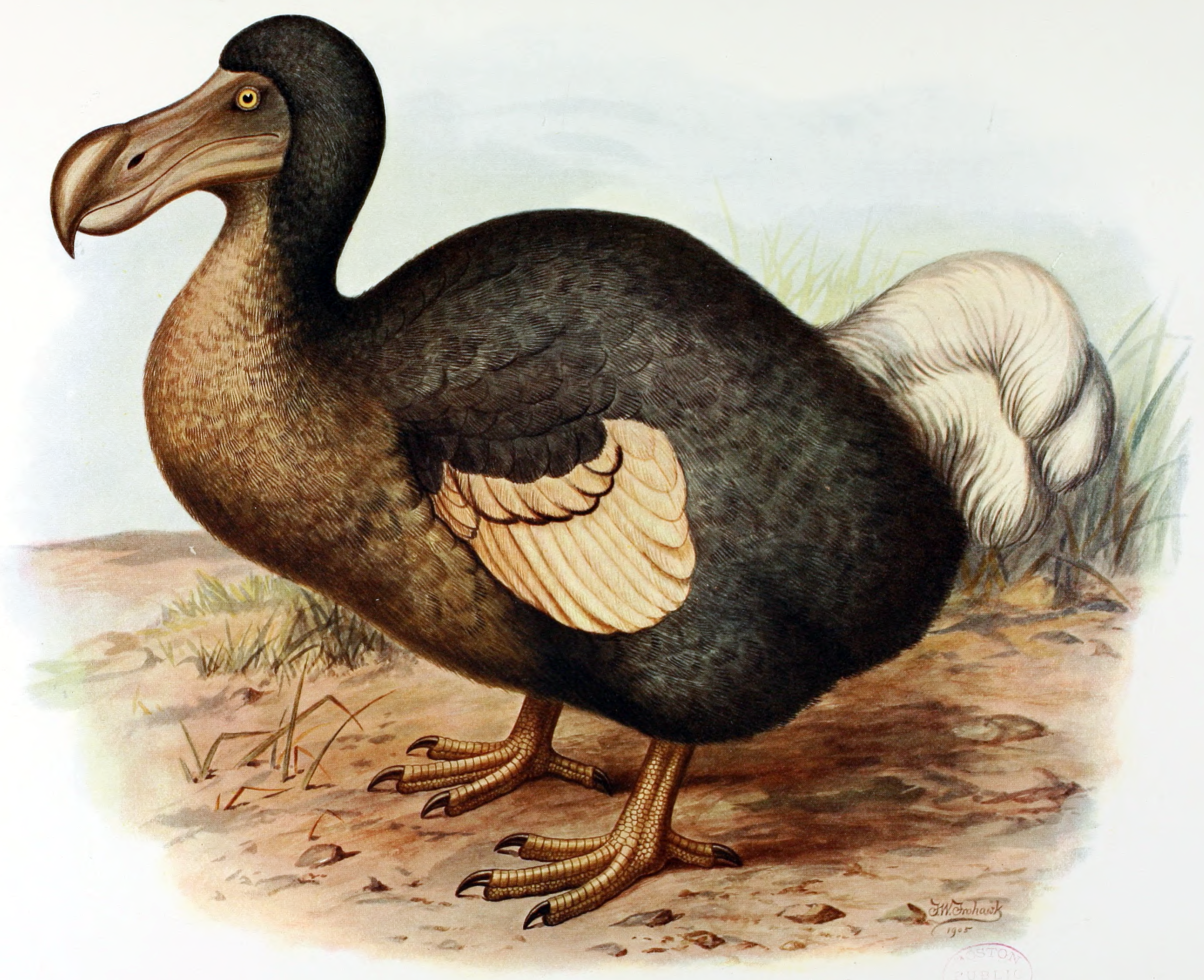
Now remember that Caloenas is in the extended dodo clade.
The dodo and its also-extinct sister taxon, the Rodrigues solitaire, form the
subfamily Raphinae.


It
also reinforces how morphologically weird Raphinae is, and how much morphological
variation the clade shows. In fact, it’s probably not surprising that it was so
hard to place the spotted green by morphology.

So,
the paper starts with a lone specimen of unknown providence and uses ancient
DNA to confirm that it was an island-hopper from Oceania and a proper species
in its own right. On top of that, they contributed to DNA extraction
methodology and say a bit about the dodo.
References
Dabney, J. et al. Complete mitochondrial genome sequence of a Middle Pleistocene cave bear reconstructed from ultrashort DNA fragments. PNAS 110, 15758–15763 (2013). Heupink, T.
H., van Grouw, H. & Lambert, D. M. The mysterious Spotted Green Pigeon and its relation to the Dodo and its kindred. BMC Evolutionary Biology 14, 136 (2014).
Pereira, S. L., Johnson, K. P., Clayton, D. H. & Baker, A. J. Mitochondrial and Nuclear DNA Sequences Support a Cretaceous Origin of Columbiformes and a Dispersal-Driven Radiation in the Paleogene. Syst Biol 56, 656–672 (2007).
The IUCN Red List of Threatened Species. Available at: http://www.iucnredlist.org/. (Accessed: 6th May 2017)
Image credits by slide
1 Specimen
https://commons.wikimedia.org/wiki/File:Caloenas_maculata_Clemency_Fisher.jpg
2 Extinct logo http://www.iucnredlist.org/
3 Sir Joseph Banks htps://upload.wikimedia.org/wikipedia/commons/6/6e/Joseph_Banks_1773_Reynolds.jpg
5 The Beatles https://commons.wikimedia.org/wiki/File:Beatles_ad_1965_just_the_beatles_crop.jpg
6 Oceania map https://upload.wikimedia.org/wikipedia/commons/thumb/4/43/Oceania%2C_broad_%28orthographic_projection%29.svg/2000px-Oceania%2C_broad_%28orthographic_projection%29.svg.png
7 Caloenas maculata reconstruction del Hoyo, J., Elliott, A., & Sargatal, J. eds. 2002. Handbook of the Birds of the World. Vol. 7. Jacamars to Woodpeckers. Lynx Edicions, Barcelona
8 Columba livida http://s0.geograph.org.uk/geophotos/01/30/95/1309587_3b1c324c.jpg Caloenas nicobarica https://upload.wikimedia.org/wikipedia/commons/3/32/Nicobar_Pigeon_RWD2.jpg Ptilinopus jambu https://upload.wikimedia.org/wikipedia/commons/4/4f/Jambu_Fruit_Dove_2010.jpg
18 Barcode https://upload.wikimedia.org/wikipedia/commons/thumb/3/3a/Example_barcode.svg/2000px-Example_barcode.svg.png
21 BLAST logo https://blast.ncbi.nlm.nih.gov/Blast.cgi
Pigeon silhouette http://commons.wikimedia.org/wiki/File:Columba_livia_Luc_Viatour.jpg
22 Maximum Likelihood tree, from Heupink et al. (2014)
23 Raphus cucullatus https://upload.wikimedia.org/wikipedia/commons/9/9b/Frohawk_Dodo.png Pezophaps solitaria https://upload.wikimedia.org/wikipedia/commons/4/47/Pezophaps_solitaria.png
29 Island in Tonga https://upload.wikimedia.org/wikipedia/commons/a/af/%27Atata_Island.JPG
2 Extinct logo http://www.iucnredlist.org/
3 Sir Joseph Banks htps://upload.wikimedia.org/wikipedia/commons/6/6e/Joseph_Banks_1773_Reynolds.jpg
5 The Beatles https://commons.wikimedia.org/wiki/File:Beatles_ad_1965_just_the_beatles_crop.jpg
6 Oceania map https://upload.wikimedia.org/wikipedia/commons/thumb/4/43/Oceania%2C_broad_%28orthographic_projection%29.svg/2000px-Oceania%2C_broad_%28orthographic_projection%29.svg.png
7 Caloenas maculata reconstruction del Hoyo, J., Elliott, A., & Sargatal, J. eds. 2002. Handbook of the Birds of the World. Vol. 7. Jacamars to Woodpeckers. Lynx Edicions, Barcelona
8 Columba livida http://s0.geograph.org.uk/geophotos/01/30/95/1309587_3b1c324c.jpg Caloenas nicobarica https://upload.wikimedia.org/wikipedia/commons/3/32/Nicobar_Pigeon_RWD2.jpg Ptilinopus jambu https://upload.wikimedia.org/wikipedia/commons/4/4f/Jambu_Fruit_Dove_2010.jpg
18 Barcode https://upload.wikimedia.org/wikipedia/commons/thumb/3/3a/Example_barcode.svg/2000px-Example_barcode.svg.png
21 BLAST logo https://blast.ncbi.nlm.nih.gov/Blast.cgi
Pigeon silhouette http://commons.wikimedia.org/wiki/File:Columba_livia_Luc_Viatour.jpg
22 Maximum Likelihood tree, from Heupink et al. (2014)
23 Raphus cucullatus https://upload.wikimedia.org/wikipedia/commons/9/9b/Frohawk_Dodo.png Pezophaps solitaria https://upload.wikimedia.org/wikipedia/commons/4/47/Pezophaps_solitaria.png
29 Island in Tonga https://upload.wikimedia.org/wikipedia/commons/a/af/%27Atata_Island.JPG
31 Didunculus strigirostris
https://upload.wikimedia.org/wikipedia/commons/c/cf/Didunculus_strigirostris.jpg
Goura scheepmakeri https://upload.wikimedia.org/wikipedia/commons/6/69/Goura_scheepmakeri_sclaterii_1_Luc_Viatour.jpg
32 Map, modified from https://upload.wikimedia.org/wikipedia/commons/thumb/f/f2/World_pacific_0001.svg/2000px-World_pacific_0001.svg.png
Goura scheepmakeri https://upload.wikimedia.org/wikipedia/commons/6/69/Goura_scheepmakeri_sclaterii_1_Luc_Viatour.jpg
32 Map, modified from https://upload.wikimedia.org/wikipedia/commons/thumb/f/f2/World_pacific_0001.svg/2000px-World_pacific_0001.svg.png